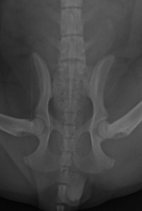
A 15-week-old spaniel pup presented to us at West Midlands Referrals with progressive ataxia (wobbliness) affecting the hind limbs. The referring vets had spotted a transitional vertebra between the lumbar and the sacral vertebrae at the bottom of the back.
Unfortunately, the clients were not insured and could not fund advanced imaging (CT scan/MRI), which would have been ideal in this case.
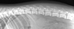
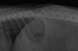
So we settled for a myelogram (done by injection of contrast just behind the skull). The contrast agent ran the full length of the spinal canal and into the sacrum in this patient. This allowed us to exclude the transitional vertebra as the cause of the current progressive wobbliness (though this vertebra may cause future issues).
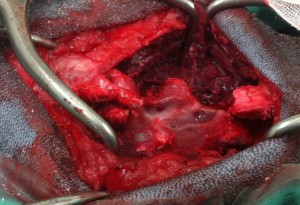
The problem at the moment was higher up the spine. A narrowing was pressing on the spinal cord between the last thoracic and the first lumbar vertebra. This related to an abnormal articular facet on the left side at this level which was situated more centrally than usual and the roof of the spinal canal was bulging down onto the spinal cord.
The area was decompressed with a high-speed air-driven spinal bur, and the patient recovered well from GA. The future should be bright, although there is always the risk of the roof of the spinal canal reforming and giving ongoing pressure to the cord at this level.
9th December 2013