There are two long bones in the lower forelimb below the elbow, the radius and the ulna. The radius and ulna's distal (lower) ends makeup part of the carpus (wrist) joint. At their proximal (top) ends, they meet the humerus forming the elbow joint.
During growth, the radius and the ulna must keep pace with each other. If growth in one outstrips growth in the other, then one is relatively too long, which can have serious consequences. The leg can end up being shorter than it should have been and can be deviated inwards or outwards. But more importantly, the elbow and carpus joints can be disrupted. The elbow is potentially the biggest problem as it is designed to be a hinge joint with a precise snug fit between the radius, ulna and humerus. The elbow is very “intolerant” of any incongruency. Degenerative joint disease results from incongruency in the elbow with depressing reliability.
Growth discrepancies between the radius and the ulna can result from injury in the first few months of life to their growth plates (the cartilaginous areas near the ends of each of the long bones from which bone growth arises). The distal (lowest) growth plate of the ulna is the growth plate that is most frequently involved with traumatic injuries of this kind. This leads to the ulna being too short in comparison with the radius In some breeds like Bassetts and Shih Tzus such “short ulna syndrome” is often present, affecting both forelimbs as a breed-related growth issue.
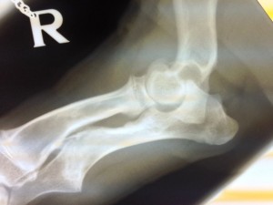
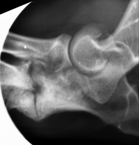
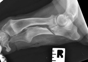
When the ulna is too short, the curved joint surface doesn’t fit well into the curved joint surface of the humerus. This makes the elbow joint incongruent. Grating and clicking can often be felt as the elbow is manipulated. The incongruency is usually obvious on radiographs taken from the side, and a “sickle” shaped tapering joint space is usually apparent. We make an oblique osteotomy (bone cut) with a surgical saw a short way below the elbow. This cut goes right the way through the ulna. This allows the triceps muscle to pull the free proximal (top) part of the ulna. The curved joint surface on the ulna can then find its position of best fit on the curved joint surface of the humerus. A gap opens up immediately between the two parts of the ulna. The parts of the ulna could be stabilised or part stabilised with metalwork, but in young patients they will usually heal naturally over a few weeks with the formation of a callus (boney lump) that can usually be felt in the weeks following on the back of the ulna over the site of the wound. Leaving the osteotomy to heal naturally has the big advantage that we aren’t dictating exactly where the ulna should end up. We leave it a big of “wiggle room” to bed itself in over a few weeks, into the position in which it is mechanically “happiest”.
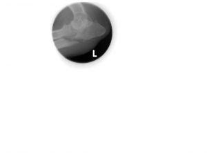
These patients will limp for a few weeks while bone healing progresses, but we monitor healing with x-rays. We can almost always take these x-rays without further sedation or anaesthesia using a hand-held x-ray generator at our Burton-on-Trent practice. When both sides need doing, we start on the worst affected side and do the other one a few weeks later.
4th November 2013