Chris joined our team in 2019 in Burton-Upon-Trent, and we’ve been pretty impressed with his skills with an endoscope! He has removed a number of grass seeds from secondary and tertiary bronchi. These cases usually present with chronic coughs and sometimes signs of grumbling infection and fever.
The trachea divides into two primary bronchi. These then divide into secondary and tertiary bronchi. The further in, the smaller the tube, the more places to check, and the harder it gets …
Removal of these foreign bodies (FB) is a challenge. It requires good equipment and a steady hand. We’ve acquired a really nice Storz video-endoscope and so cool grasping tools that fit down the working channel in the scope, so the whole enterprise of removing these objects is very much like a surreal video game.
CT beforehand can be useful to get an idea of where they are located in the lungs, though they do tend to follow set paths when they inhaled, often ending up in the caudal lung lobes, and usually flying in like darts, so the backwards pointing awns can make removal a challenge.
Time spent fiddling these pesky grass seeds and awns out though, is time very well spent and makes a huge difference to the quality of lives of these dogs.

Grass in a cat’s bronchus
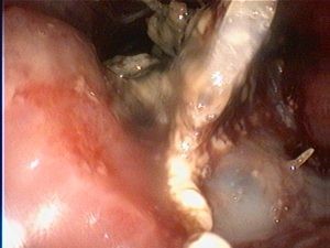
Metal graspers closing around the grass so it can be grasped and pulled out
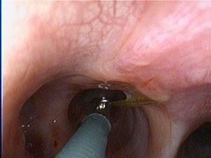
Grass seed awn in a dog’s tertiary bronchus
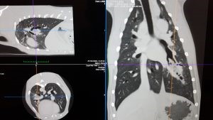
CT imaging beforehand is useful for locating the FB
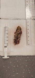
The grass seed awn, removed from a dog’s bronchus
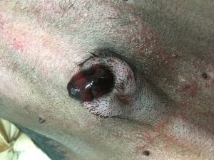
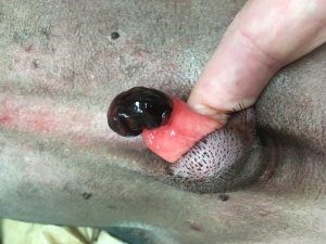
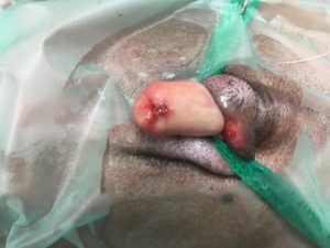
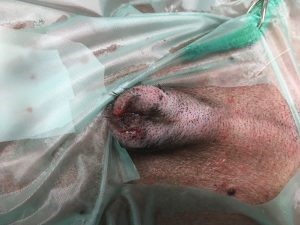
A 7-year-old neutered male Staffie presented with urethral prolapse which was resected and sutured around a catheter. The catheter was then removed. The prognosis is generally very good for these cases.