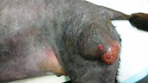
Benson, a middle-aged Labrador retriever, had a skin tumour on his left thigh which had unfortunately grown to a large size before it was investigated.
At West Midlands Veterinary Referrals we were asked to look at Benson while we were visiting the referring practice to operate on another case there. Benson’s mass was biopsied with sedation and local anaesthetic while the owner waited. The excised tissue samples were submitted to an external lab and the results confirmed that the mass was a mast cell tumour, a common kind of skin tumour that derives from a population of granular cells that are involved with inflammatory processes and wounds. Furthermore, the tumour was graded as “intermediate” and the potential local invasion of surrounding tissues was a consideration.
Excision of the tumour was planned. Tumours are removed with a “margin” of healthy tissue to ensure that all of the tumour is removed. The more locally aggressive/invasive the tumour is, the bigger the margin that is required. It is important to get well under the tumour to ensure a deep margin, as well as just cutting widely around it. In this case we wanted 2cm margins, and we removed substantial parts of one of the three hamstring muscles to ensure an adequate deep margin. Removing this muscle has no significant long term adverse effect as the other two hamstring muscles “bulk up” to take the extra load. But removing substantial hamstring muscle was expected to make the surgery painful. Closure of the resulting wound, which would inevitably be very large, was planned. We intended to use skin flaps derived from the groin and possibly even from below his abdomen if more tissue was required.
Benson was anaesthetised and the desired 2cm margin of excision was marked out from the edges of the mass. As the incision was made in front of the leading edge of the mass, inflammation and haemorrhage into tissue under the skin was identified. We could not know for sure that this wasn’t in fact tumour excision, and so the plan had to be changed. If the tumour had invaded at the point where we had incised and if we had then created skin flaps, we would have risked spreading tumour cells along his body wall.
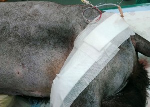
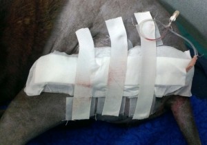
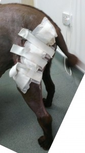
The wound before closure looks far too gory to picture here, but it was a real challenge to get it together without flaps. We did manage to close it without further elevation of skin flaps by using deep tension sutures and by using a Velcro/elastic system of skin stretchers to support the skin and surface tissues while they healed. We expected that this would be even more painful post-operatively and so Benson’s analgesia protocol needed beefing up before he was recovered from anaesthetic. He was already on methadone and non-steroidal anti-inflammatory analgesia. We added in ketamine as a constant rate infusion in his drip, and his wound was closed over a fenestrated wound irrigation cannula that allowed us to infiltrate local anaesthetic into the wound several times in the early postoperative period. This cannula was placed so that it exited the skin very close to the main wound. This meant that any contamination of fresh tissue by any tumour cells migrating along the drain would be insignificant. The elastic skin stretchers were tightened periodically in the hours following surgery as they pulled in more superficial tissue, taking the strain off the sutured wound.
Whatever they made Benson out of, it was seriously tough stuff. The analgesia protocol was robust, but even so, this dog’s pain threshold was simply off the scale. He trotted around on the leg quite happily post op, wagged his tail, and left his wound completely alone.
A Courier submitted the excised mass to an external laboratory for examination. The suspect area in front of the mass was marked to allow the pathologist to pay particular attention to it. The lab reported that the desired margins of 2cm had been achieved or exceeded everywhere around and under the tumour except for the area that we had been concerned about intra-operatively. We had in fact achieved clear margins here too, but these margins were down to a little under 1cm in places. The inflammation/haemorrhage had probably been the result of the inflammatory substances that mast cells release, but fortunately wasn’t the result of local invasion.
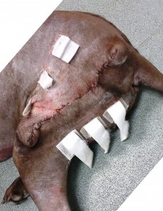
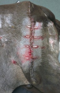
The wound held together well. Benson had a nibble at his wound about 10 days post-op. The Velcro/elastic was coming off the skin at this point as the skin it was glued to aged and was shed. So the elastic / Velcro was removed. Under sedation, we reknotted the end of the skin suture that he had started to unravel, and placed staples to reinforce the rest of the wound. Four stent sutures – sutures through short pieces of plastic tubing to stop them from pulling through – were placed to reinforce the wound edges.
Further special testing of the excised mass is in hand so that we’ll be well placed to add in medical therapy/chemotherapy if this proves necessary.
17th August 2013