This case is a favourite – a horrendous fracture, a challenging surgery, planning with CT advanced imaging, a lovely cat, patient and committed owners, and a great outcome. What’s not to like.
Stanley had an RTA and his pelvis was, to coin a technical expression, mashed. His right ischium (the wing of the pelvis) was in several bits and these were rotated every which way. One was rammed up the middle of the pelvis and will have battered the lumbosacral nerve trunk, the major innervation of the hind limb.
The referring vets x-rays were very good. Even so, I struggled to see how the bits would fit back together.
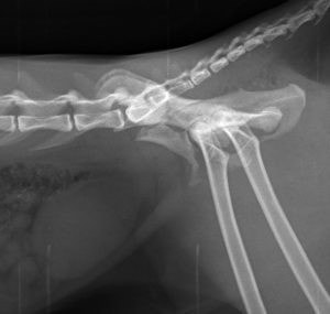
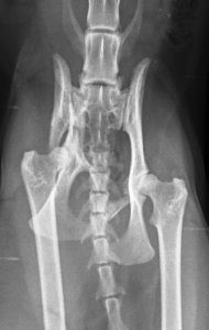
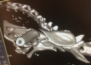
Time to cheat and use the CT scanner. These are “stills” from the CT, but with the software, you can spin the pelvis any way you want and “look” at it from any angle. Very handy in planning.
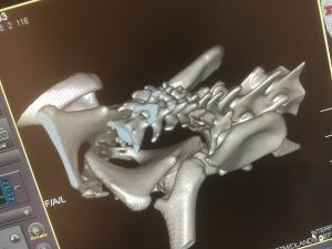
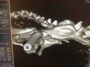
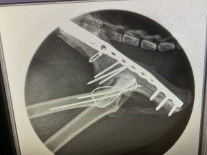
Access required cutting the top of the femur, subsequently fixed with pins and wire, reconstructing the pelvis with more pins and then sliding a locking plate under the sciatic nerve to buttress the repair.
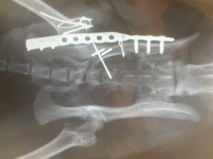
One pin was a little long, but all in all, it came together pretty well.
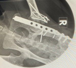
Gradually, in the weeks that followed surgery, the nerve trunk recovered, millimetre by millimetre, and he progressed from a cat dragging a knuckly leg around to one who could walk normally 3-4 months later
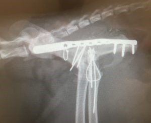
These images were taken conscious – he let us hold him up while we took the x-ray views with a handheld machine. The bone is healing nicely. The implants stay where they are unless they cause issues in the future, in which case out they will come. He’s signed off for rechecks now and has his life back.
