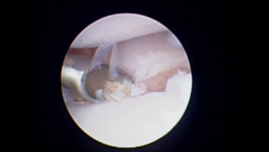
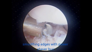
Placement of a “camera” into joints – most commonly the elbow or the shoulder – allows for inspection with magnification and for some procedures to be minimally invasive without a fully open surgical procedure.
We see a lot of Labradors and Retrievers for scoping with the medial coronoid disease. The medial coronoid process is a spur on the upper end of the ulna, one of the bones in the forearm. This boney process, and the cartilage that overlies it, can fragment. Removal of these fragments is usually possible arthroscopically, with tools and powered burs inserted into the joint through small holes and via access tubes. This approach is less invasive and offers the benefit of magnification compared to a traditional open surgical approach to achieve the same ends.
Most patients are improved in the short-mid term, though regretfully, the intervention does not improve a minority of cases. There is an inevitable progression of osteoarthritis (degenerative joint disease, DJD) in these cases regardless.
6th June 2016


