Rupture of the cranial cruciate ligament is one of the commonest problems that is referred to us. Rupture of this ligament can be partial or complete and the cartilages in the knee can be damaged because of the instability that results.
We operate on several of these cases per week. These injuries can either be treated with one of a group of techniques using a suture around the outside of the joint to give support while periarticular fibrosis or scarring develops, or more commonly by using one of a number of bone-cutting techniques to change the way that the knee works to help the patient to cope with a ruptured cranial cruciate ligament. The procedure that we most commonly recommend is tibial tuberosity advancement (TTA). The TTA allows dogs to use the knee much more comfortably despite the ruptured cruciate ligament. The fixed prices that we charge are so competitive that we have had cases coming to us from as far afield as Southampton and Southend. The savings that these clients have made in so doing has easily justified the travel.
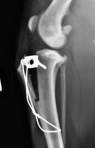
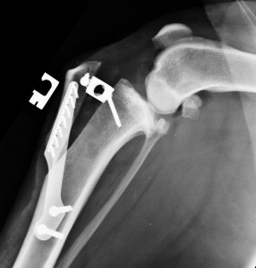
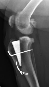
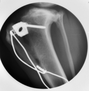
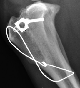
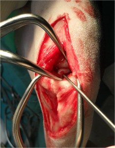
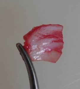
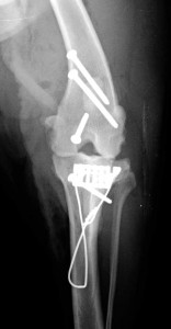
A cut is made in the bone of the tibia below the knee to create a gap. A cage spacer is inserted to maintain the gap. The cut bone can then be stabilised in a number of ways while the gap fills in with new bone. The original technique uses a fork and plate for stabilisation. A variation in technique that has been developed in the last couple of years involves leaving a small part of the bone cut intact. A wire is commonly then used to add extra stability. The cage spacers are made of titanium and a recent variant of the TTA technique uses an expanded titanium foam wedge instead of the titanium open cage.
Recommended aftercare following surgery for cranial cruciate ligament rupture, and an information sheet on cruciate ligament disease can be found in the section of fact sheets, located within the section for owners on this website.
Typically patients treated with TTA are taking significant weight through the operated knee within a few days of surgery. The patients are kept on a lead with gradually increasing periods of lead exercise in the weeks following surgery. Hydrotherapy typically starts around 3-4 weeks post-operatively. We take check X-rays to confirm satisfactory healing of the bone at about 8 weeks post-operatively. We do this while the owners wait, usually using our hand-held X-ray generator, and usually without even the need for sedation. Under our fixed scheme, we make no further charges for any of our follow up checks or for any follow up radiography that we do.
Free exercise can be allowed once bone healing is satisfactorily progressed, typically from about 8 weeks post-operatively.
29th December 2013
